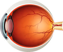
The eye is a highly complex organ responsible for the sense of sight. It allows us to perceive the world around us by detecting light and converting it into electrical signals that the brain processes into images. Here’s a detailed breakdown of the anatomy and functioning of the eye:
1. External Parts of the Eye:
These are the structures you can see on the outside of the eye.
- Eyebrow: Helps to protect the eyes from sweat and debris. It also plays a role in non-verbal communication by showing emotions.
- Eyelids: The eyelids protect the eye from foreign bodies and bright lights. They also help distribute tears across the surface of the eye to keep it moist. The blink reflex helps remove dust and keeps the eye’s surface lubricated.
- Eyelashes: These act as sensors, alerting you to potential threats like dust, and help to protect the eyes from particles.
- Sclera: The white part of the eye, the sclera is a tough, protective outer layer made of connective tissue. It maintains the eye’s shape and serves as an attachment point for the muscles that move the eye.
2. Structures Inside the Eye:
Cornea:
- Structure: Transparent and dome-shaped, the cornea is the outermost layer of the eye.
- Function: It serves as the first refractive surface that bends (refracts) incoming light to help focus it on the retina. It also plays a significant role in protecting the eye from dirt, germs, and other environmental hazards.
Anterior Chamber:
- Structure: Located between the cornea and the iris, this is filled with a watery fluid known as aqueous humor.
- Function: The aqueous humor nourishes the cornea and lens and helps maintain intraocular pressure, which helps the eye maintain its shape.
Iris:
- Structure: The colored part of the eye, composed of muscles and pigments.
- Function: The iris controls the size of the pupil (the black circular opening in the center of the iris) by contracting and relaxing. This helps regulate the amount of light that enters the eye. It is responsible for adjusting the pupil to bright or dim light.
Pupil:
- Structure: The circular black hole in the center of the iris.
- Function: The pupil dilates (opens) in low light to allow more light in and constricts (closes) in bright light to limit the light entering the eye. The size of the pupil is controlled by the iris.
Lens:
- Structure: A transparent, flexible, and biconvex (curved on both sides) structure located directly behind the iris and pupil.
- Function: The lens focuses light onto the retina by changing shape (a process known as accommodation). For near objects, the lens becomes more curved; for distant objects, it flattens out. This fine-tuning allows us to focus clearly on objects at various distances.
Vitreous Body (Humor):

- Structure: A clear, gel-like substance that fills the space between the lens and retina.
- Function: The vitreous body helps maintain the shape of the eye and supports the retina by holding it in place against the back of the eye. It also provides structural support.
3. Inner Eye Structures:
Retina:
- Structure: A thin, light-sensitive layer at the back of the eye. The retina contains photoreceptor cells called rods and cones.
- Rods are sensitive to low light and are responsible for black-and-white vision.
- Cones are responsible for color vision and function best in bright light.
- Function: The retina detects light and converts it into electrical signals. These signals are sent to the brain via the optic nerve for processing.
Macula:
- Structure: A small, central area of the retina.
- Function: The macula is responsible for sharp, detailed central vision. It contains a high concentration of cones and allows us to focus on objects directly in front of us.
Fovea:
- Structure: Located in the center of the macula, the fovea is a small pit.
- Function: The fovea contains only cones and is the area of the retina responsible for the highest visual acuity (sharpness of vision). This is where the eye focuses when you look directly at something.
Optic Nerve:
- Structure: A bundle of nerve fibers that extends from the retina to the brain.
- Function: The optic nerve transmits electrical signals from the retina to the brain, where they are interpreted as visual images.
4. Supporting Structures and Fluids:
Optic Disc (Blind Spot):
- Structure: The area where the optic nerve leaves the eye.
- Function: There are no photoreceptors (rods or cones) in this area, which creates a “blind spot.” However, this is not noticeable under normal circumstances because the brain fills in the gaps in vision.
Aqueous Humor:
- Structure: A watery fluid found in the anterior chamber (between the cornea and the lens).
- Function: The aqueous humor nourishes the cornea and lens and maintains intraocular pressure, which helps the eye retain its shape. It is continuously produced and drains out of the eye to maintain pressure balance.
Ciliary Body:
- Structure: A circular muscle structure surrounding the lens.
- Function: The ciliary body produces the aqueous humor and contains muscles that control the shape of the lens for focusing.
5. How the Eye Works:
- Light enters the eye through the cornea and pupil.
- The cornea and lens work together to focus the light onto the retina.
- The light-sensitive rods and cones in the retina convert the light into electrical signals.
- The electrical signals travel through the optic nerve to the visual cortex in the brain, where they are interpreted as images.
6. Common Eye Conditions and Disorders:
Here are some common problems that affect the eye:
- Refractive Errors: Issues like nearsightedness (myopia), farsightedness (hyperopia), and astigmatism, which affect how light is focused on the retina, causing blurry vision.
- Presbyopia: The age-related loss of the ability to focus on close objects, often due to a stiffening of the lens.
- Cataracts: A clouding of the lens, leading to blurry vision, which usually occurs with aging.
- Glaucoma: Damage to the optic nerve, often caused by increased eye pressure, which can lead to vision loss.
- Macular Degeneration: A disease that affects the macula (the central part of the retina), leading to the loss of central vision.
- Diabetic Retinopathy: Damage to the retina caused by diabetes, which can lead to vision loss.
- Retinal Detachment: A medical emergency where the retina pulls away from its normal position, leading to vision loss.
The eye is a remarkable organ that allows us to perceive the world in vivid detail. Understanding its intricate structure and functions highlights the complexity of sight and its importance to our daily lives.

Leave a Reply